Joint, Muscle & Rheumatology
Knee Pain
Focused on the knee — the commonest painful weight-bearing joint — covering causes from osteoarthritis to meniscus and ligament problems, and how knee pain is assessed and managed.
Overview
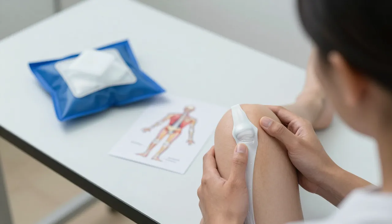
The knee is the joint most often troubled by pain, largely because it carries the body’s weight through every step. Knee pain may come from the joint itself (arthritis, meniscus, cartilage), the tissues around it (ligaments, tendons, bursae), or be referred from the hip or back. This page focuses on the knee; the broader symptom picture is on the Joint Pain page, and the wear-and-tear disease on the Osteoarthritis page.
Common causes
- Osteoarthritis — the commonest cause, especially with age and excess weight.
- Meniscus (cartilage) tears — often with twisting or squatting, sometimes with locking or giving way.
- Ligament injuries — such as an ACL tear, often after sport.
- Patellofemoral (front-of-knee) pain — common in younger active people.
- Bursitis and tendon problems — such as housemaid’s knee or jumper’s knee.
- Gout or pseudogout — sudden hot, swollen knee (see Gout).
- Infection — a hot, swollen knee with fever, needs urgent assessment.
What the history and examination show
The story matters: gradual worsening with use and stiffness suggests osteoarthritis; a twist or fall with swelling within hours suggests a meniscus or ligament injury; a sudden hot knee at night suggests gout. Examination checks for swelling, warmth, tenderness, range of movement, stability of the ligaments, and how the kneecap tracks. Weight is an important factor, because every kilogram multiplies the load on the knee with each step (see Obesity & Weight Management).
Evaluation and investigations
An X-ray of the knee is the first-line test, showing joint-space narrowing and osteoarthritis changes. Blood tests (inflammatory markers, uric acid) are added when inflammation or gout is suspected. MRI is arranged when a meniscus, ligament or cartilage problem needs detail beyond the X-ray.
Management overview
Most knee pain is managed without surgery — weight management, activity modification, quadriceps-strengthening exercise, physical therapy, and short careful courses of simple pain relief. A flare of osteoarthritis may be settled with a targeted injection in selected cases. Structural injuries (meniscus, ligament) and infections need orthopaedic assessment. See Osteoarthritis for the disease-specific plan.
Lifestyle and activity
Low-impact activity — walking, cycling, swimming — keeps the knee moving while protecting the cartilage. Strengthening the thigh muscles (quadriceps) is one of the most effective measures for knee arthritis, because strong muscles absorb load. Avoiding prolonged squatting and kneeling helps where these aggravate the pain.
Warning signs — seek prompt care
- A hot, red, very swollen knee with fever.
- Knee pain after a serious injury with inability to bear weight.
- Sudden swelling with locking or inability to straighten the knee.
How we help
Dr. Nayak identifies the source of your knee pain, orders the right imaging and tests, and builds a non-surgical plan — with orthopaedic referral when a structural injury or infection is found. Consultations are available in-clinic in Bhubaneswar and over teleconsultation.
Related conditions and patient concerns
Explore more
Related conditions
Explore more
Related services
Take action now
Schedule your consultation today with Dr. Nayak
Don't wait to get the healthcare you deserve. Book your appointment in just a few clicks.